Cusabio Cell adhesion Recombinants
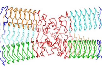
Abstract Due to the biocompatibility and biodegradability, as well as the mechanical properties of the fibres, spider silk has become an attractive material for researchers […]

Abstract Due to the biocompatibility and biodegradability, as well as the mechanical properties of the fibres, spider silk has become an attractive material for researchers […]
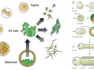
Abstract The current state of knowledge strongly supports the view that development and differentiation are a function of variable gene activity. The crucial area of […]
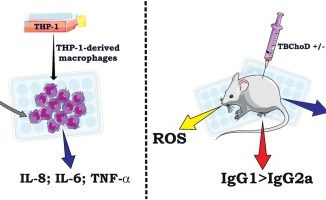
Bottom The current limitations of conventional BCG vaccines highlight the importance in the development of new and effective vaccines against tuberculosis (TB). The use of […]

Low move sequencing has been proposed as an economical different to genotyping arrays to determine genetic variants that affect multifactorial traits in people. For frequent […]

Genotyping of polymorphic chromosomal inversions in malaria vectors comparable to An. coluzzii Coetzee & Wilkerson is necessary, each as a result of they trigger cryptic […]

Phosphatidylinositol-4,5-bisphosphate 3-kinase catalytic subunit alpha (PIK3CA) mutation has a prevalence of roughly 2% in lung adenocarcinoma. However, instances presenting a PIK3CA mutation alone have hardly […]

Genome enhancing in mice utilizing both classical approaches like homologous recombination or CRISPR/Cas9 has been reported to harbor off track results (insertion/deletion, body shifts or […]

Epidemiological and molecular organic information counsel that β-Human Papillomavirus (HPV) could cause epithelial skin tumours, however the relationship stays unclear. A brand new method to […]

The interpretation of quick tandem repeat (STR) profiles will be difficult when, for instance, alleles are masked due to allele sharing amongst contributors and/or when […]
Scientists from EPFL and INRAE have developed a new approach to create artificial proteins, the results of which in vivo, as vaccines, have shown promise. […]
Copyright © 2024 | WordPress Theme by MH Themes